Breadcrumbs
Case of the Month: December 2024
Clinical history
An 84-year-old woman was referred to a dermatology clinic for an ulcerating nodule on her left cheek. She has no significant past medical history. The dermatologist took a biopsy of the nodule and wrote on the requisition: fast-growing lesion; rule out squamous cell carcinoma, basal cell carcinoma, melanoma, Merkel cell carcinoma.
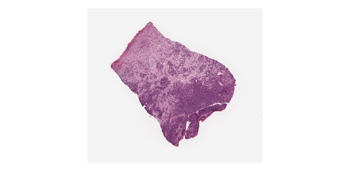
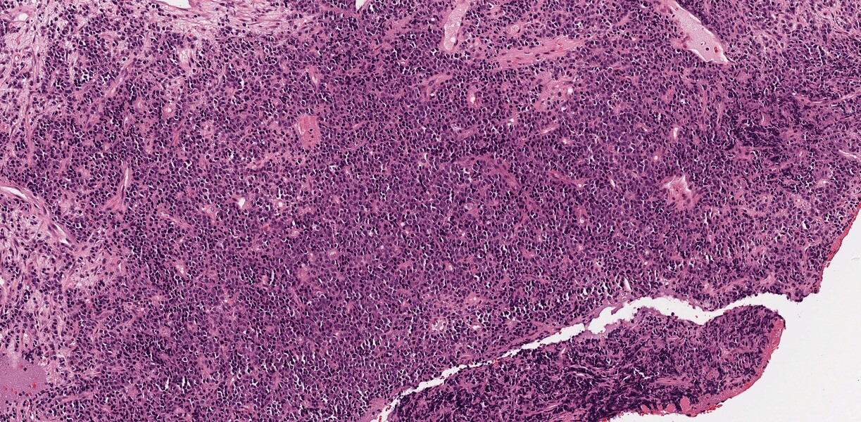
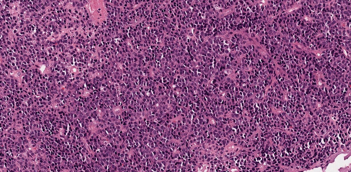
Review the slide image “H&E”. This is a biopsy of a skin lesion. At low power, you should expect to see normal skin; however, the normal skin architecture has been completely effaced, which should prompt you to be suspicious of a neoplastic process. On low power, you can see sheets of monotonous appearing cells. On high power, you can see the cells are medium-sized, have high nuclear: cytoplasmic ratio and hyperchromatic to fine pale chromatin. There are occasional mitotic figures and apoptotic bodies.
Questions:
- What are the main categories of cancer by cell of origin (i.e. what is the type of cancer arising from an epithelial cell, etc)?
- Which immunohistochemical markers are positive?
- Which cancer types can we exclude from our differential diagnosis list?
How to participate
If you are a medical student at the University of Toronto, you can also:
If you do, you are in for a chance to win a $100 gift card! (UofT Med students only).

H&E 2x

H&E 10x

H&E 20x
If you are a current medical student at U of T, you can register to join the January case of the month lecture online or in person and have a chance to win a $100 gift card!
You can also join the Special Interest Group in Laboratory Medicine to learn more about Laboratory Medicine.
Laboratory Medicine and the study of disease for medical students

